Skip to content
  • Stanford Medicine
  • •
  • Otolaryngology — Head & Neck Surgery
Oto Surgery Atlas Logo Oto Surgery Atlas Logo Oto Surgery Atlas Logo
  • Atlas
  • About This Atlas
Fundamentals of Ear SurgeryTori Lewis Fibonacci Web Studio2020-08-19T21:21:11-07:00
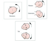
The three variables for positioning of the head in ear microsurgery: rotation, tilt, and flexion–extension.

Patient Positioning

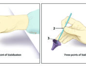
Incorrect hand position for ear microsurgery, which is less stable and limits visibility. The correct hand position has three-point stabilization.

Microinstrument Hand Positioning

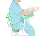
A sizable fraction of ear microsurgeons develop back problems over time. This can be mitigated by using proper posture during microsurgery. This involves upright posture and good lumbar support as shown here. Many surgeons use rests to enhance stability and alleviate arm fatigue.

Ergonomics

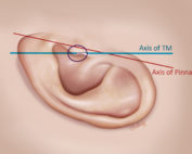
The pinna tilts posterior about 15 degrees with respect to the axis of the malleus.

Postauricular Incision

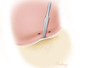
Commencing elevation of the ear canal flap (skin and periosteum) from Henle’s spine. Note the cribriform region just posterior to the spine.

Postauricular Canal Flap Design

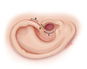
There are three segments to an endaural incision: circumferential, intercartilaginous (between the helical crus and the tragus), and the vertical limb. It is less commonly used today compared to the postauricular incision due to the visibility of the scar and limitation of exposure posteriorly.

Endaural Incision

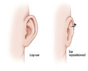
In closing a postauricular incision following tympanomastoid procedures, the pinna may rotate outward and develop a cosmetically undesirable prominence (i.e., lop ear). This is especially the case in children due to the flexibility of their cartilage.

Incision Closure

  • Welcome to Otologic Surgery Atlas
  • Surgical Anatomy of the Ear
    • Overview of Temporal Bone
    • External Ear
    • Middle Ear & Mastoid
    • Eustachian Tube
    • Auditory System
    • Vestibular System
    • Internal Auditory Canal and Cerebellopontine Angle
    • Ear Tumor Anatomy
  • Fundamentals of Ear Surgery
    • Patient Positioning
    • Microinstrument Hand Positioning
    • Ergonomics
    • Postauricular Incision
    • Postauricular Canal Flap Design
    • Endaural Incision
    • Incision Closure
  • External Ear
    • Otoplasty
    • Microtia Repair
    • Repair of Congenital Aural Atresia
    • Postauricular Meatoplasty
    • Endaural Meatoplasty
    • Exostoses of the Ear Canal
    • Anterior Canalplasty
    • Preauricular Cyst Excision
  • Stapes Surgery
    • Overview
    • Exposure
    • Stapedotomy
    • Stapedectomy
    • Challenges
    • Deficient Incus
    • Revision Stapes Surgery
    • Tympanosclerosis
  • Tympanoplasty
    • Tympanic Membrane Perforations
    • Fascia Harvesting
    • Tragal Cartilage Harvesting
    • Medial Graft Tympanoplasty
    • Anterior Perforation Repair
    • Lateral Graft Tympanoplasty
    • Butterfly Tympanoplasty
    • Eustachian Tuboplasty
  • Ossiculoplasty
    • Patterns of Ossicular Deficiency
    • Autologous Ossiculoplasty
    • Prosthetic Ossiculoplasty
    • Ossiculoplasty Placement Technique
    • Trans-facial Recess Ossiculoplasty
    • Medialized Malleus
    • Staging Ossiculoplasty
  • Mastoidectomy
    • Intact Canal Wall Mastoidectomy
    • Anatomical Variation in Mastoidectomy
    • Complications of Mastoidectomy
    • Facial Recess Approach
    • The Facial Nerve in Mastoidectomy
    • Mastoid Obliteration
  • Cholesteatoma
    • Growth Patterns of Cholesteatoma
    • Canal Wall Down Mastoidectomy
    • Microdissection of Cholesteatoma
    • Retrograde Technique
    • Epitympanum Repair
    • The Facial Nerve in Cholesteatoma
    • Sinus Tympani & Facial Recess in Cholesteatoma
    • Anterior Epitympanic Cholesteatoma
    • Semicircular Canal Fistula in Cholesteatoma
    • Ossicles in Cholesteatoma
    • Congenital Cholesteatoma
  • Facial Nerve
    • Anatomy of the Facial Nerve
    • Repair of the Facial Nerve
    • Hypoglossal–Facial Anastomosis for Facial Reanimation
    • Hypoglossal-Trigeminal-Facial Anastomoses
    • Gracilis Microvascular Facial Reanimation
    • Static and Dynamic Slings for Facial Reanimation
  • Vestibular Surgery
    • Endolymphatic Sac Surgery
    • Labyrinthectomy
    • Superior Semicircular Canal Dehiscence
  • Cochlear Implants
    • Cochlear Implant Surgery
    • Cochlear Implant Surgical Variations
  • Temporal Bone Fractures, Encephaloceles, and Cerebrospinal Fluid Leaks
    • Temporal Bone Fractures
    • Encephalocoeles
    • Cerebrospinal Fluid Leaks
    • Ear Canal Closure
  • Temporal Bone Resection
    • Lateral Temporal Bone Resection
    • Extended Temporal Bone Resection
  • Petrous Apex
    • Petrous Apicotomy
    • Petrous Apicectomy
    • Transsphenoidal Approach to Petrous Apex
  • Pulsatile Tinnitus
    • Pulsatile Tinnitus of Dural Sinus Origin
    • Pulsatile Tinnitus of Carotid and Jugular Bulb Origin
  • Appendix: Educational Handouts for Patients

© Stanford Medicine • Privacy Policy • Terms of Use